Understanding the inner universe with Cryo-Electron Tomography
Feb 03 2022
Author: Julika Radecke on behalf of Diamond Light Source Ltd
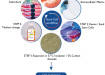
Everything we do from moving a finger to thinking and memory formation, triggers a signalling cascade in the brain and body. Neurotransmitters (NTs) are released from the synapse - the nerve terminal between neurons where information is passed from one neuron to the next - into the synaptic cleft, the 20-30 nm space between neurons, and onto the beginning of the neighbouring cell which is called the postsynapse (Figure 2).
If movement is involved, eventually the signal from the brain will reach a designated muscle cell instructing it to contract which could lead, for example, to the movement of a finger. Information about a new position of the finger will be immediately sent back to the brain from receptor neurons to check if intended results are achieved or if any correction is required.
If we want to understand how the brain and nervous system is affected by diseases, injury, or mental health conditions, its fine structure and function must be studied. High resolution microscopy techniques play a crucial role in this quest; they are important for visualising the fine details needed to develop treatments.
In the past, brain cells were studied after fixation involving dehydration and staining by chemicals, to preserve a snapshot of the grand structure of neurons in a particular state. However, this meant fine details were lost, such as small protein filaments in synapses that provide communication between the cells. These structures, called tethers and connectors, connect one vesicle to another. It is important to investigate small proteins like these because they are often affected in neurodegenerative diseases.
低温电子断层扫描(cryo-ET)允许我们研究独特的对象,如神经元和连接器的详细信息,而不需要化学染色和脱水。
低温em和低温et是如何工作的?
Cryo-ET is a branch of cryo-Electron Microscopy (cryo-EM). The name of the method underlines that samples are studied at very low temperatures, normally at -196°C. At this temperature water, which makes over 90% of content in all living organisms, can exist in amorphous non-crystalline form, also found in comets in deep space.
During cryo-EM sample preparation, the water in the sample is turned into amorphous ice (called vitrification) by rapid plunging of the sample into liquid ethane, which is cooled down by liquid nitrogen to about -172°C. Biological samples are vitrified so rapidly that ice crystal formation is prevented, and structural integrity preserved as if they are still in their natural environment. The samples are said to be preserved in a frozen-hydrated state, since no drying occurs during sample preparation and study, for vitreous ice can be kept in the electron microscope vacuum at temperatures below -156°C.
在cryo-ET中,通过结合不同角度采集的样本的多个投影,可以建立一个独特样本的三维(3D)模型。电子可以像医学断层扫描中的x射线一样通过样本,但由于电子是带电的,电磁透镜可以用来创建一个非常小的物体的详细放大图像,使我们能够探索亚细胞分子结构的宇宙(在[1]回顾)。
Moreover, by subjecting cells to various treatments with chemical and physical stimuli, and then taking snapshots after the treatment, one can potentially study tiny changes. This allows understanding of how the cells function, enabling the creation of a dynamic, 4-dimensional (4D) model of cellular processes.
Cryo-ET produces nanometer-scale information in the form of high-resolution (~1–4 nm) 3D views of samples, typically biological macromolecules, and cells. The technique is ideally suited to study thicker cellular specimen in their native cellular context, but optimal sample thickness is below 300 nm.
For unlocking details of thicker samples, thinning by Focused Ion Beam milling in a dual beam Scanning Electron Microscope (SEM) has been invented (cryo-FIB SEM). A thin section of the cell can be used for building 3D models using cryo-ET [1]. Application of this technique by scientists at the UK’s electron Bio-Imaging Centre (eBIC) at Diamond Light Source helped to shed light on the life cycle of SARS-CoV-2 at the onset of the global Covid-19 pandemic (Figure 1) [2].
Learning cryo-EM and cryo-ET techniques for neuroscience
我在德国慕尼黑的马克斯·普朗克心理学研究所攻读理学硕士学位后,对高分辨率显微技术产生了兴趣。我想更彻底地了解大脑的结构,所以我研究了哪些显微镜可以实现更高的分辨率,并发现了令人兴奋的新技术,如低温em,一种高分辨率成像技术,在2017年获得了诺贝尔奖。
2012年,我搬到瑞士伯尔尼的解剖研究所攻读博士学位,并做了一段时间的博士后研究。博士学位是通过冷冻- et(另一项当时仍处于起步阶段的技术)研究突触小泡在突触小体(捏断神经末梢)中的胞外分泌。
我真的很喜欢细胞断层扫描,因为在细胞内有许多有趣的东西吸引眼球,让你想知道它们可能是什么。这让我想研究细胞内的所有过程,但不幸的是,时间有限!
Learning the amazing cryo-ET technique, developing, and improving ways to achieve millisecond resolution with an atomizer as originally used by Berriman and Unwin [3], and analysing the tomograms for smallest changes on the synaptic vesicle and the opposing active zone plasma membrane was very intriguing. I also had the opportunity to expand my knowledge in protein biochemistry, cell culture and related sample preparation for cryo-EM processing, as well as learning cryo-FIB. Ultimately, my aim was to unravel ongoing cellular processes studied by cryo-light and electron microscopy.
To delve deeper into neuroscience, I obtained an early career postdoctoral research grant from the Swiss National Science Foundation to join a lab in Copenhagen, Denmark, in 2017. I initially learned electrophysiology and chromaffin cell preparation but then moved on to primary astrocyte and neuron cell culture.
Understanding neurotransmitter release (synaptic vesicle (SV) exocytosis or signal transduction) in healthy neurons - which proteins are involved and what happens during release - are the necessary basics before researching how diseases affect brain functionality.
The synapse contains synaptic vesicles which are filled with signalling molecules. Our research looked at functional aspects of synaptic vesicle exocytosis to understand which proteins are involved and what happens when certain proteins are knocked out through a gene deletion.
Attempting to grow neurons on EM grids presented a challenge
最终,我成功地建立了一种在EM网格上生长神经元的方法,这种方法也使许多薄的功能性突触也具有突触后(图2)。在适当的护理和条件下,在网格上生长初级神经元可以很简单,但在网格上获得一个足够薄的功能突触(指突触、间隙和突触后)却不容易。
Studying the brain, Sars-CoV-2 vaccine research and supporting eBIC microscope users
2019年,我开始在eBIC担任电子显微镜高级支持科学家。我以前的经验,特别是冷冻et的经验,为我担任这个角色做好了准备。
Around 70% of my time is focused on support to UK and worldwide users who send us samples. I help them with experiment set-up such as data acquisition strategies, sample preparation, and advise on what to improve. We also work on other projects as well as our own.
For example, after the Covid-19 pandemic started we dedicated all our resources to help investigate the Covid-19 virus to beat the disease. We investigated the viral spike protein and worked on cellular samples to see what the very first vaccines being developed were doing in the cells.
采用单粒子低温em技术对从病毒本身分离的刺突蛋白进行研究,揭示了刺突蛋白的结构[4]和低温et,用于细胞研究。通过了解尖刺是如何进入细胞的,以及它使用的结构,可以开发特定的药物来干扰该结构,以阻止细胞进入[2]。这是辅以其他先进的x射线成像和衍射技术,我们在钻石。结合软x射线成像与低温et和系列聚焦离子束扫描电镜,可以描述SARS-CoV-2感染的详细图像[Mendonca et al[2]]。x射线晶体学提供了高分辨率的结构数据,可用于加速药物发现过程,并在Diamond成功地用于理解,例如,担忧的变体如何逃脱自然免疫或疫苗诱导免疫[参考文献- Stuart等[5]的细胞论文]。
我在eBIC的另外30%的工作是研究自己的项目,通过研究大脑结构和功能之间的关系来更好地理解大脑。在一个健康的系统中了解这一点,可以改善我们如何了解受疾病或精神健康状况影响的系统,以开发治疗方法。我的助手是一名在工业界学习的学生,他们研究初级神经元培养,并获得x光片来研究突触内的小蛋白。
我们在eBIC采用了一系列最新的高分辨率显微镜技术。我发现尽可能多地了解显微镜的技术细节是很有趣的。和Diamond的工程师一起解决显微镜的故障,以及学习如何解决一些更容易的问题,都是非常棒的。最近,我发现自己在一个Talos Arctica TEM的盒子里,试图找到一个特定的电缆,需要拔掉插头来重置连接!
High resolution microscopy - what experience and qualifications are needed?
有动力去学习显微镜的物理/光学特性对更深入地理解显微镜和如何操作它们是有用的。
就我而言,我是带着普通生物学背景来到我的研究领域,专攻神经科学的。我在生物学本科期间参加了为期两周的显微镜强化课程,在此期间我学习了如何使用光学显微镜(LMs)。我进展到使用共聚焦LM在我的硕士项目,从那里移动到EM在我的博士期间。
现在我给一个三年级的理学学士教授EM和神经科学。她非常有上进心,非常渴望学习如何操作显微镜和如何生长神经元的所有细节。
在戴蒙德,有几个x射线设备可以用于研究同一个问题,但在不同的规模,从成像更大的体积使用断层扫描或相干成像,或研究细胞内的化学使用光谱技术。在戴蒙德大学和校园内的研究机构都有很多合作的机会。由于工作环境舒适、灵活,因此在电磁领域以及其他领域和技术上有很大的发展和进步空间。要亲身体验这些技巧并发现更多,在Diamond有很多职业选择,比如申请行业一年、暑期实习或博士学位。
Ultimately, one needs to be curious and motivated to learn new things, alongside a willingness to adapt ideas and opinions. With the right mindset there’s always a role that suits.
References
1. Turk, M. and Baumeister, W. (2020), The promise and the challenges of cryo-electron tomography. FEBS Lett, 594: 3243-3261, https://doi.org/10.1002/1873-3468.13948
2. Mendonça L, Howe A, Gilchrist JB, Sun D, Knight ML, Zanetti-Domingues LC, Bateman B, Krebs AS, Chen L, Radecke J, Sheng Y, Li VD, Ni T, Kounatidis I, Koronfel MA, Szynkiewicz M, Harkiolaki M, Martin-Fernandez ML, James W, Zhang P. Correlative multi-scale cryo-imaging unveils SARS-CoV-2 assembly and egress. Nat Commun 12, 4629 (2021), https://doi.org/10.1038/s41467-021-24887-y
3. John Berriman, Nigel Unwin, Analysis of transient structures by cryo-microscopy combined with rapid mixing of spray droplets, Ultramicroscopy, Volume 56, Issue 4, 1994, Pages 241-252, ISSN 0304-3991, https://doi.org/10.1016/0304-3991(94)90012-4
4. Dejnirattisai, W., Zhou, D., Ginn, H. M., Duyvesteyn, H. M. E., Supasa, P., Case, J. B., Zhao, Y., Walter, T. S., Mentzer, A. J., Liu, C., Wang, B., Paesen, G. C., Slon-Campos, J., López-Camacho, C., Kafai, N. M., Bailey, A. L., Chen, R. E., Ying, B., Thompson, C., …, Radecke J, … Screaton, G. R. (2021). The antigenic anatomy of SARS-CoV-2 receptor binding domain, Cell, Volume 184, Issue 8, 2021, Pages 2183-2200.e22, ISSN 0092-8674, https://doi.org/10.1016/j.cell.2021.02.032 (https://www.sciencedirect.com/science/article/pii/S009286742100221X)
5. Stuart et al : Reduced neutralization of SARS-CoV-2 B.1.617 by vaccine and convalescent serum DOI: 10.1016/j.cell.2021.06.020 June 2021; The antibody response to SARS-CoV-2 Beta underscores the antigenic distance to other variants DOI: 10.1016/j.chom.2021.11.013 Nov 2021; SARS-CoV-2 Omicron-B.1.1.529 leads to widespread escape from neutralizing antibody responses DOI: 10.1016/j.cell.2021.12.046 Jan 2022
Electron Bio-Imaging Centre (eBIC): https://www.diamond.ac.uk/Instruments/Biological-Cryo-Imaging/eBIC.html
Career options at Diamond Light Source: https://www.diamond.ac.uk/Careers.html
Figure 1 Image adapted from [2] Nat Commun 12, 4629 (2021), https://doi.org/10.1038/s41467-021-24887-y. Reproduced in accordance with the Creative Commons license: http://creativecommons.org/licenses/by/4.0/.
Digital Edition
Lab Asia 29.4 - August 2022
August 2022
In This Edition Chromatography - Automated Sample Preparation:The Missing Hyphen to Hypernation - New Low Volume Air Sampler for PFAS Analysis - Analytical Intelligence Starts with the Samp...
View all digital editions


Events
ACS National Meeting & Expo, Fall 2022
Aug 21 2022Chicago, IL, USA & Online
Aug 22 2022Frankfurt, Germany
Aug 27 2022Maastricht, Netherlands
Aug 28 2022Lisbon, Portugal
Aug 31 2022Singapore